The Science
Every living cell emits light. Cancer changes the pattern. HelioFlux reads that change at 5-10 million cells, long before imaging, blood tests, or symptoms.
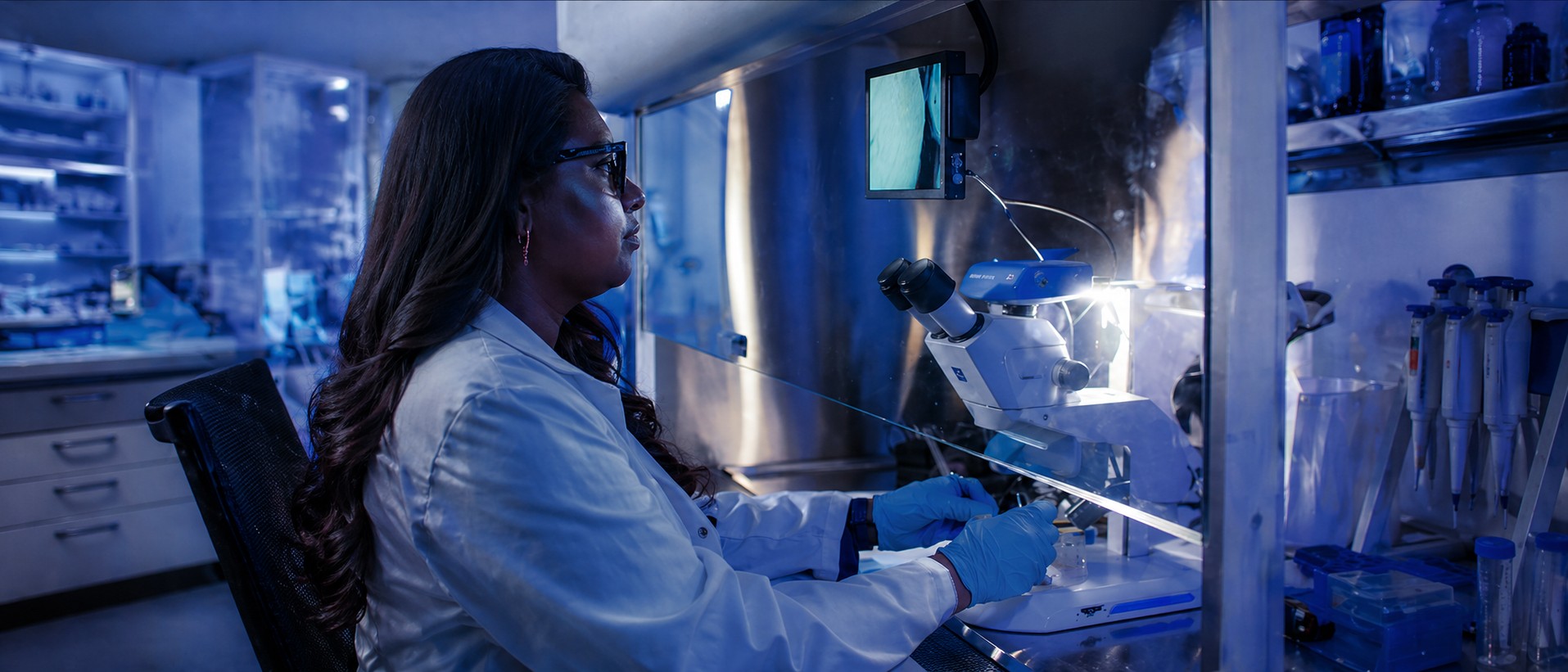
How It Works
A QSense sensor is placed against the skin. It counts individual photons — the faint light your cells are always emitting — at single-photon sensitivity.
The raw signal feeds into LuminAI, our NVIDIA-powered software. It runs spectral analysis in real time, comparing the emission fingerprint against a validated database of cancer and healthy-tissue signatures.
In about 15 minutes, the clinician has a result. No biopsy. No blood draw. No radiation. No waiting for the lab.
The Physics
Ultra-weak photon emissions (UPEs) are a byproduct of oxidative metabolism — the chemical reactions that power every cell. They are real, measurable, and carry information about the cell's internal state. Healthy cells produce a characteristic emission profile. Cancer cells do not.
The difference lies in the spectral shape, not brightness. Malignant cells emit photons at shifted wavelengths, with a frequency signature around ~20Hz that does not appear in healthy tissue. This signature was discovered by Dr. Nirosha Murugan across a decade of peer-reviewed research.
LuminAI applies spectral power density (SPD) analysis to extract this fingerprint from raw photon data. The result is a biomarker classification — malignant or not — grounded in physics, not symptoms, imaging, or blood chemistry.
Detection Threshold
A tumor at 5-10 million cells is about 2 millimeters across. Too small to feel. Invisible on a scan. By the time it reaches 1 billion cells, roughly a centimeter, medicine can finally see it. That growth took 1.5 to 2 years. HelioFlux is built to find it first.
Our Pilot
Every year, millions of patients leave a dermatologist's office with an unanswered question: is this mole dangerous? The doctor has two options. Cut it out and send it to a lab, or say "let's watch it." Most biopsies come back benign. The ones that don't sometimes come back too late.
HelioFlux gives clinicians a third option. A 15-minute scan reads the photon signature of a suspicious lesion and returns a result in the office. No cutting. No waiting. No guessing.
Skin is where we start because the clinical pathway is clear: the dermatologist already has the patient, already suspects something, and already faces a binary decision. HelioFlux plugs into that moment without changing the workflow.
Once we validate the technology on melanoma, the same platform expands. Brain cancer. Breast cancer. Eventually, a full-body scan that reads every tissue type at once.
Preclinical Evidence
In 2020, Dr. Murugan's lab published results from 47 live animal models injected with melanoma cells. Whole-body photon emissions detected the cancer within 24 hours of injection — 18 days before tumors were physically detectable. Final discriminant accuracy: 90%.
This is the in vivo foundation for HelioFlux's human skin pilot. Same detection principle. Same spectral signature. Now built into a clinical-grade device.
Read the study (Murugan et al., 2020) →Published Research
Only 5 of 200+ cancer types have a recommended screening protocol. HelioFlux is designed to change that.